
Hot Topics and Position Statements
Topics of Interest and Talking Points
The topics below reflect areas where radiologists may encounter questions, conflicting information, or misconceptions from patients, referring providers, or others. These resources are intended to equip radiologists with clear, evidence-based, informed talking points to help navigate those conversations and provide appropriate clinical context.
Abstract: Screening mammography has been proven to reduce the mortality from breast cancer by approximately 30%, however, it is less sensitive in women with dense breast tissue and certain risk groups. Supplemental screening may be considered based on the patient’s risk level and breast density. In all women, digital breast tomosynthesis improves screening sensitivity. Average-risk women with heterogeneously dense tissue may also benefit from breast MRI, abbreviated breast MRI (AB-MRI) or breast ultrasound (US). In intermediate-risk women with nondense tissue, breast MRI and ABMRI may be appropriate. In intermediate-risk women with heterogeneously dense and extremely dense tissue, breast MRI and AB-MRI are usually appropriate, whereas US and contrast-enhanced mammography (CEM) may be appropriate. Breast MRI or ABMRI is usually appropriate in all high-risk women, regardless of density. Screening breast US or CEM could be considered in this population.
- Thermography is not FDA approved as an independent breast cancer screening tool and can only be used as adjunctive. Breast Cancer Screening: Thermogram No Substitute for Mammogram | FDA
- Multiple large breast cancer screening trials on thermography were stopped early or showed no benefit over mammography.
- Thermography can miss obvious cancers and has a low sensitivity (~50%) compared to mammography, ultrasound, and MRI.
- Thermography is not recommended by the Society of Breast Imaging, American College of Radiology, or American Society of Breast Surgeons.
- Patients with questionable thermography findings should undergo diagnostic mammography, ultrasound, or MRI to determine management.
- Thermography is not typically covered by insurance and results in out-of-pocket expenses for patients, unlike screening mammography.
Abstract: Ductal carcinoma in situ (DCIS) accounts for approximately 20% of diagnosed breast cancer. It is important to understand which imaging studies are appropriate in patients with a new diagnosis or history of DCIS. Initial imaging for a new diagnosis of DCIS, consists of diagnostic mammography and/or tomosynthesis, whereas breast ultrasound and breast MRI may be appropriate as complementary examinations. Routine surveillance with annual mammography and/or tomosynthesis is recommended to detect an in-breast recurrence or a new primary breast cancer in women who have completed breast conservation therapy for DCIS, and breast MRI may be appropriate. Advanced technologies such as contrast mammography or molecular breast imaging are usually not appropriate. In a patient with a history of mastectomy for DCIS, routine surveillance for ipsilateral recurrence with imaging is usually not appropriate. There is no role for imaging of the axilla in patients with known DCIS with or without microinvasion.
- Mammography uses ionizing radiation. Ionizing radiation is carcinogenic at high doses. Breast MRI and ultrasound do not use ionizing radiation.
- The carcinogenic risk of low-dose radiation (effective doses below 100 mSv) is negligible. No radiation-induced cancers have been observed at such low doses. All radiology imaging exams, including mammograms, are well below this 100 mSv threshold.
- The effective dose from digital mammographic screening is approximately 0.5 mSv. Since annual background radiation in the United States is approximately 3.1 mSv, digital screening mammograms are equivalent to 8 weeks of natural background radiation.
- The significant mortality benefit of diagnosing breast cancer at earlier stages substantially outweighs the small, theoretical carcinogenic risk of radiation from mammography.
- Tomosynthesis adds a radiation dose equal or up to 1.5x conventional digital mammography. Thus, combination digital mammography + tomosynthesis results in an overall radiation dose double that of conventional digital mammography alone. Using synthetic 2D mammography reduces the radiation dose by almost half (i.e., overall dose is similar to conventional digital mammography alone).
- Thyroid shields are unnecessary during mammograms because the thyroid dose from scatter radiation is exceedingly small. For perspective, the effective dose to the thyroid from a digital screening mammogram is 0.13 microSv, which is equivalent to 30 minutes of natural background radiation.
Abstract: Given that 20% to 40% of women who have percutaneous breast biopsy subsequently undergo breast surgery, knowledge of imaging women with a history of benign (including high-risk) disease or breast cancer is important. For women who had surgery for nonmalignant pathology, the surveillance recommendations are determined by their overall risk. Higher-than-average risk women with a history of benign surgery may require screening mammography starting at an earlier age before 40 and may benefit from screening MRI. For women with breast cancer who have undergone initial excision and have positive margins, imaging with diagnostic mammography or MRI can sometimes guide additional surgical planning. Women who have completed breast conservation therapy for cancer should get annual mammography and may benefit from the addition of MRI or ultrasound to their surveillance regimen.
Abstract: Breast pain (or tenderness) is a common symptom, experienced by up to 80% of women at some point in their lives. Fortunately, it is rarely associated with breast cancer. However, breast pain remains a common cause of referral for diagnostic breast imaging evaluation. Appropriate workup depends on the nature and focality of the pain, as well as the age of the patient. Imaging evaluation is usually not indicated if the pain is cyclic or nonfocal. For focal, noncyclic pain, imaging may be appropriate, mainly for reassurance and to identify treatable causes. Ultrasound can be the initial examination used to evaluate women under 30 with focal, noncyclic breast pain; for women 30 and older, diagnostic mammography, digital breast tomosynthesis, and ultrasound may all serve as appropriate initial examinations. However, even in the setting of focal, noncyclic pain, cancer as an etiology is rare.
https://www.jacr.org/article/S1546-1440(18)31156-6/fulltextPatient and Provider Resources
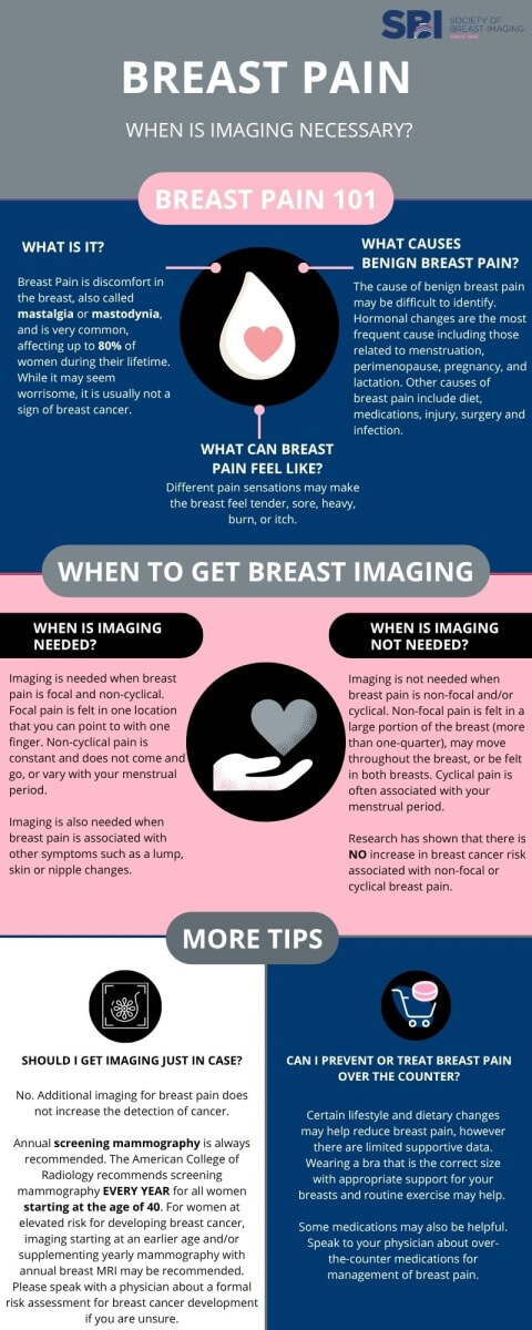
Patients often have questions about breast cancer screening, so we’ve created a handout with answers to frequently asked questions. You can access the provider handout here and the patient handout here.
For providers, we’ve also developed a printable poster to display in offices or share on digital platforms. Access on our flickr page here.
Abstract: For asymptomatic patients with saline implants, no imaging is recommended. If there is suspected saline implant rupture, ultrasound is usually appropriate, though saline implant rupture is often clinically evident. For asymptomatic patients with silicone implants, the FDA recommends that patients have an initial ultrasound or MRI examination without contrast 5 to 6 years after initial silicone implant surgery and then every 2 to 3 years thereafter. In a patient with silicone implants and suspected implant complication, MRI without contrast is usually appropriate and ultrasound and/or mammography may be appropriate, depending on age. In a patient with unexplained axillary adenopathy with current or prior silicone breast implants, ultrasound and/or mammography are usually appropriate, depending on age. In the setting of a patient with breast implants of any type and suspected implant-associated malignancy, ultrasound or MRI without and with contrast is usually appropriate as initial imaging test.
Abstract: Although the majority of male breast problems are benign with gynecomastia as the most common etiology, men with breast symptoms and their referring providers are typically concerned about whether or not it is due to breast cancer. If the differentiation between benign disease and breast cancer cannot be made on the basis of clinical findings, or if the clinical presentation is suspicious, imaging is indicated. The panel recommends the following approach to breast imaging in symptomatic men. In men with clinical findings consistent with gynecomastia or pseudogynecomastia, no imaging is routinely recommended. If an indeterminate breast mass is identified, the initial recommended imaging study is ultrasound in men younger than age 25, and mammography or digital breast tomosynthesis in men age 25 and older. If physical examination is suspicious for a male breast cancer, mammography or digital breast tomosynthesis is recommended irrespective of patient age.
Abstract: This publication reviews the current evidence supporting the imaging approach of the axilla in various scenarios with broad differential diagnosis ranging from inflammatory to malignant etiologies. Controversies on the management of axillary adenopathy results in disagreement on the appropriate axillary imaging tests. Ultrasound is often the appropriate initial imaging test in several clinical scenarios. Clinical information (such as age, physical examinations, risk factors) and concurrent complete breast evaluation with mammogram, tomosynthesis, or MRI impact the type of initial imaging test for the axilla. Several impactful clinical trials demonstrated that selected patient populations can receive sentinel lymph node biopsy instead of axillary lymph node dissection with similar overall survival, and sentinel lymph node biopsy is a safe alternative as the nodal staging procedure for clinically node negative patients or even for some node positive patients with limited nodal tumor burden. This approach is not universally accepted, which adversely affect the type of imaging tests considered appropriate for axilla. This document is focused on the initial imaging of the axilla in various scenarios, with the understanding that concurrent or subsequent additional tests may also be performed for the breast.
Abstract: This article provides evidence-based guidance for imaging lactating women across screening, diagnostic, and staging scenarios. Imaging should not be deferred because of lactation; mammography with digital breast tomosynthesis, targeted ultrasound, and contrast-enhanced MRI have defined roles based on age, presenting complaint, and cancer risk and may be used similarly to nonlactating patients. Physiologic changes during lactation can alter breast appearance and complicate clinical and imaging assessment. Gadolinium contrast and most nuclear medicine procedures result in negligible infant exposure and do not require interruption of breastfeeding. Image-guided core biopsy and aspiration are safe and effective, with rare lactation-specific risks, which should be discussed during consent. Recommendations aim to standardize care, minimize diagnostic delay, preserve breastfeeding when possible, and support multidisciplinary decision-making. These guidelines inform clinicians, radiologists, and multidisciplinary teams in routine practice.
Abstract: There are physiologic and structural changes of the breast that occur during pregnancy that can make the clinical examination and imaging more challenging. Pregnancy-associated breast cancer (PABC) is uncommon but increasing as more women postpone childbearing. PABC can present clinically as a palpable lump, focal pain, nipple discharge, or diffuse breast enlargement. Mammography is safe to perform during pregnancy. Both high- and normal-risk women should continue their routine screening mammography examinations during pregnancy. Diagnostic mammography and/or ultrasound is tailored to evaluate the clinical symptoms and for locoregional staging of newly diagnosed PABC. Breast MRI is not advocated for screening or diagnostic evaluation of the breast during pregnancy due to unknown safety of gadolinium exposure to the fetus.
Abstract: Palpable masses in women are the most common symptom associated with breast cancer. This document reviews and evaluates the current evidence for imaging recommendations of palpable masses in women less than 30 to over 40 years of age. There is also a review of several different scenarios and recommendations after initial imaging. Ultrasound is usually the appropriate initial imaging for women under 30 years of age. If ultrasound findings are suspicious or highly suggestive of malignancy (BIRADS 4 or 5), it is usually appropriate to continue with diagnostic tomosynthesis or mammography with image-guided biopsy. No further imaging is recommended if the ultrasound is benign or negative. The patient under 30 years of age with a probably benign ultrasound may undergo further imaging; however, the clinical scenario plays a role in the decision to biopsy. For women between 30 to 39 years of age, ultrasound, diagnostic mammography, tomosynthesis, and ultrasound are usually appropriate. Diagnostic mammography and tomosynthesis are the appropriate initial imaging for women 40 years of age or older, as ultrasound may be appropriate if the patient had a negative mammogram within 6 months of presentation or immediately after mammography findings are suspicious or highly suggestive of malignancy. If the diagnostic mammogram, tomosynthesis, and ultrasound findings are probably benign, no further imaging is necessary unless the clinical scenario indicates a biopsy.
https://www.jacr.org/article/S1546-1440(23)00184-9/abstractAbstract: The type of nipple discharge dictates the appropriate imaging study. Physiologic nipple discharge is common and does not require diagnostic imaging. Pathologic nipple discharge in women, men, and transgender patients necessitates breast imaging. Evidence-based guidelines were used to evaluate breast imaging modalities for appropriateness based on patient age and gender. For an adult female or male 40 years of age or greater, mammography or digital breast tomosynthesis (DBT) is performed initially. Breast ultrasound is usually performed at the same time with rare exception. For males or females 30 to 39 years of age, mammography/DBT or breast ultrasound is performed based on institutional preference and individual patient considerations. For young women less than 30 years of age, ultrasound is performed first with mammography/DBT added if there are suspicious findings or if the patient is at elevated lifetime risk for developing breast cancer. There is a high incidence of breast cancer in males with pathologic discharge. Men 25 years and older should be evaluated using mammography/DBT and ultrasound added when indicted. In transfeminine (male-to-female) patients, mammography/DBT and ultrasound are useful due to the increased incidence of breast cancer. The ACR Appropriateness Criteria are evidence-based guidelines for specific clinical conditions that are reviewed annually by a multidisciplinary expert panel. The guideline development and revision process support the systematic analysis of the medical literature from peer-reviewed journals. Established methodology principles such as Grading of Recommendations Assessment, Development, and Evaluation or GRADE are adapted to evaluate the evidence. The RAND/UCLA Appropriateness Method User Manual provides the methodology to determine the appropriateness of imaging and treatment procedures for specific clinical scenarios. In those instances in which peer-reviewed literature is lacking or equivocal, experts may be the primary evidentiary source available to formulate a recommendation.
https://www.jacr.org/article/S1546-1440(22)00654-8/fulltextAbstract: Breast cancer screening recommendations for transgender and gender nonconforming individuals are based on the sex assigned at birth, risk factors, and use of exogenous hormones. Insufficient evidence exists to determine whether transgender people undergoing hormone therapy have an overall lower, average, or higher risk of developing breast cancer compared to birth-sex controls. Furthermore, there are no longitudinal studies evaluating the efficacy of breast cancer screening in the transgender population. In the absence of definitive data, current evidence is based on data extrapolated from cisgender studies and a limited number of cohort studies and case reports published on the transgender community.
Abstract: Routine screening substantially reduces the risk of mortality and morbidity of breast cancer with early detection. Multiple different imaging modalities may be used to screen for breast cancer. Screening recommendations differ based on an individual’s risk of developing breast cancer. Numerous factors contribute to breast cancer risk, which is frequently divided into three major categories: average, intermediate, and high risk. For patients assigned female at birth with native breast tissue, mammography and digital breast tomosynthesis are recommended for breast cancer screening in all risk categories. In high-risk patients, screening with breast MRI is recommended starting as early as 25 to 30 years of age and mammography and digital breast tomosynthesis with a variable starting age between 25 and 40 years of age, depending on the type of risk.
Abstract: Imaging plays a vital role in managing patients undergoing neoadjuvant chemotherapy, as treatment decisions rely heavily on accurate assessment of response to therapy. This document provides evidence-based guidelines for imaging breast cancer before, during, and after initiation of neoadjuvant chemotherapy.
Abstract: Mastectomy may be performed to treat breast cancer or as a prophylactic approach in women with a high risk of developing breast cancer. In addition, mastectomies may be performed with or without reconstruction. Reconstruction approaches differ and may be autologous, involving a transfer of tissue (skin, subcutaneous fat, and muscle) from other parts of the body to the chest wall. Reconstruction may also involve implants. Implant reconstruction may occur as a single procedure or as multistep procedures with initial use of an adjustable tissue expander allowing the mastectomy tissues to be stretched without compromising blood supply. Ultimately, a full-volume implant will be placed. Reconstructions with a combination of autologous and implant reconstruction may also be performed. Other techniques such as autologous fat grafting may be used to refine both implant and flap-based reconstruction. This review of imaging in the setting of mastectomy with or without reconstruction summarizes the literature and makes recommendations based on available evidence.
Abstract: Early detection decreases breast cancer death. The ACR recommends annual screening beginning at age 40 for women of average risk and earlier and/or more intensive screening for women at higher-than-average risk. For most women at higher-than-average risk, the supplemental screening method of choice is breast MRI. Women with genetics-based increased risk, those with a calculated lifetime risk of 20% or more, and those exposed to chest radiation at young ages are recommended to undergo MRI surveillance starting at ages 25 to 30 and annual mammography (with a variable starting age between 25 and 40, depending on the type of risk). Mutation carriers can delay mammographic screening until age 40 if annual screening breast MRI is performed as recommended. Women diagnosed with breast cancer before age 50 or with personal histories of breast cancer and dense breasts should undergo annual supplemental breast MRI. Others with personal histories, and those with atypia at biopsy, should strongly consider MRI screening, especially if other risk factors are present. For women with dense breasts who desire supplemental screening, breast MRI is recommended. For those who qualify for but cannot undergo breast MRI, contrast-enhanced mammography or ultrasound could be considered. All women should undergo risk assessment by age 25, especially Black women and women of Ashkenazi Jewish heritage, so that those at higher-than-average risk can be identified and appropriate screening initiated.
White Papers
Information
Access a variety of informative and popular resources designed to assist breast imaging radiologists in providing quality care.
Breast Screening Leadership Group Resources
- ACR Appropriateness Criteria
- Breast Screening Leadership Group & Course
- Benefits of Screening Mammography: Data from Population Service Screening
- Breast Cancer Screening: Understanding the Randomized Controlled Trials
- Limitations of the Canadian National Breast Screening Studies
- Overdiagnosis
- Screening in the 40-49 Age Group
Legislative Updates
- How to Advocate at Your State Legislature
- ACR Capitol Hill Day and Beyond
- Legislative and Regulatory Update 2024
- Legislative and Regulatory Update: An Event 2023 Thus Far!
- Radvocacy: 2022 Year in Review
Position Statements and Recommendations
2026
2025
2023
2022
2021
The Canadian Society of Breast Imaging provides the following position statements and guidelines.
The European Society of Breast Imaging provides the following position statements and guidelines.